High Temperature Raman Measurement Techniques
Raman spectroscopy is an effective technique for collecting molecular information in various process and experimental conditions. Many phase changes and reactions of interest happen in high temperature and/or high-pressure conditions. Raman measurements can be used to collect vital information in these extreme conditions and it can even be simultaneously used for temperature determinations (1) (2). As Raman is a non-contact technique, measurements can be carried out from a distance reducing risk of thermal damage to measurement devices.
A significant challenge with high temperature Raman measurements is interference caused by thermal emission. This interference can easily obscure lower intensity Raman responses and decrease signal to noise ratios in temperatures exceeding 1300 K or even 1800 K.
Most common methods for limiting thermal interference with Raman measurements include:
-
- Time-resolved techniques
- UV excitation techniques
- Spatial filtering
Time-resolved techniques
In high temperature measurements, the measured intensity is a sum of Raman signal and thermal emission (fluorescence not discussed here). The aim is to keep the detected Raman scattering to thermal emission ratio as high as possible for optimal results. Pulsed time-resolved techniques aim to do this by using energy dense laser pulses and by only collecting data during the resulting pulses originating from the sample. The figure below compares continuous wave and pulsed data acquisition with high temperature applications. This comparison also applies to other continuous interferences e.g. ambient light.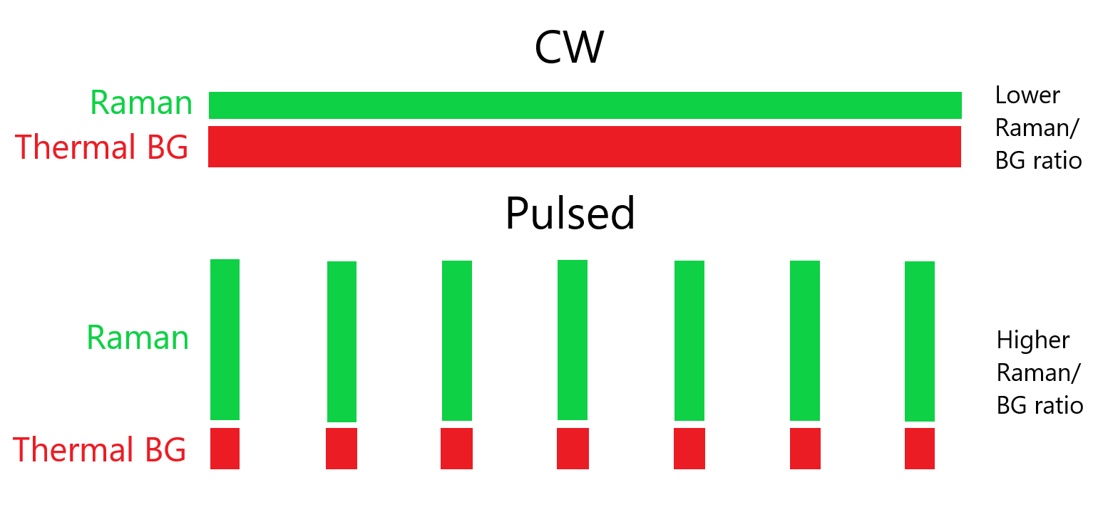
Whereas continuous wave techniques collect information continuously, pulsed systems only collect data during short pulses. As these pulses have a high peak power and the detector is not collecting information between pulses, the resulting Raman to thermal background ratio is high. High power pulsed lasers may promote sample degradation, but this may not be an issue with samples that will have to be able to withstand very high temperatures.
Improved Raman to thermal ratios are achieved when the laser pulses have high energies and when the pulses are short (time-wise). This requires very fast detectors that are in sync with the pulsed lasers. Fast detection and improved high temperature results have been achieved in the nanosecond scale using CCD and ICCD detection (3) (4) and modern SPAD based techniques can even reach 100 – 200 ps time-gating for optimal results (5) (6).
An additional benefit of pulsed time-resolved techniques is that the thermal background can be measured right before a pulse. This way the background measurement can be repeated often, and the measurement result will not include any Raman responses as the measurement takes place between excitation pulses. This background measurement result can then be used to further subtract thermal influence.
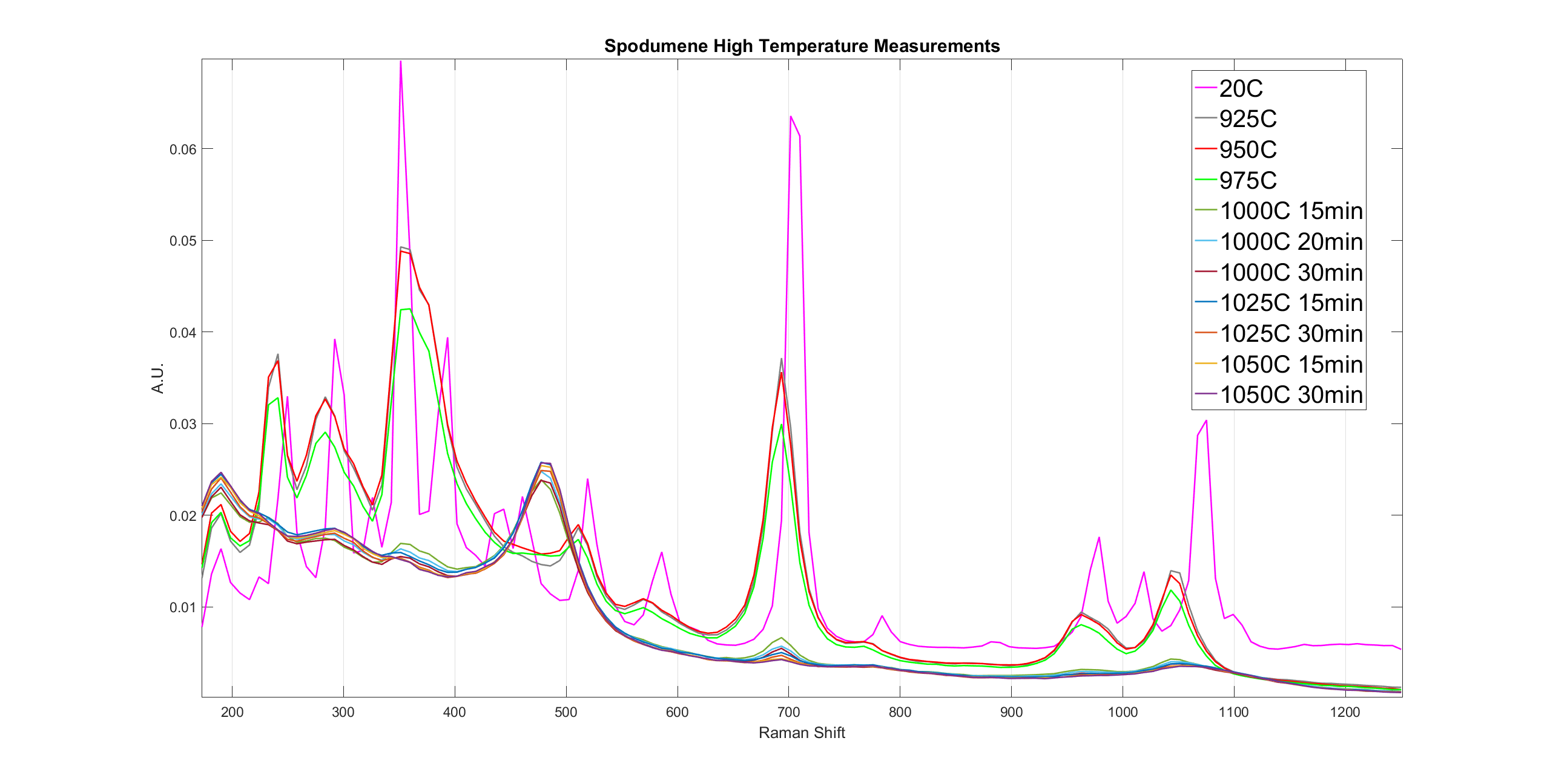
As an example, shown below are spectra from a spodumene calcination process measured with a time-gated SPAD spectrometer in >1200 K temperatures. The phase change from α-spodumene to β-spodumene is clearly visible. For more information on this high temperature application please see our spodumene calcination case study by downloading it below.

For measurements carried out in even higher temperatures (>1800K), see our previous post regarding high-temperature Raman measurements at Shanghai University.
UV excitation techniques
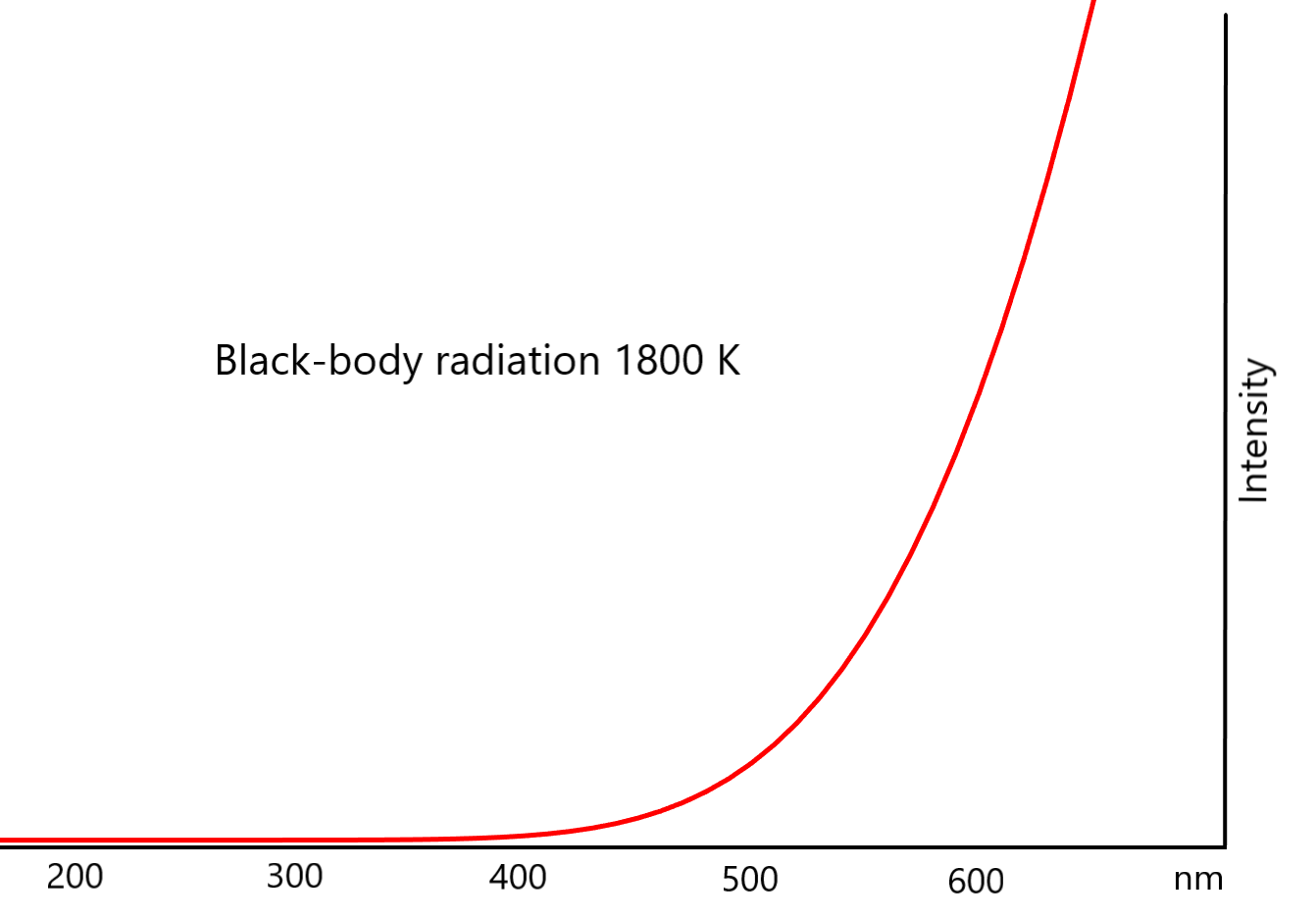
Using UV excitation can produce promising results as the short wavelength excitation shifts the Raman scattering away from the thermal emission peak (3) (7) (8). The figure below depicts black body radiation at 1800 K and it shows why collecting Raman scattering at lower wavelengths can be beneficial for avoiding thermal emission interference.
Excitation in the UV range may also produce Raman scattering intensity benefits due to resonance effects (9). However, UV excitation may induce luminescence interference (3) and strong absorption in the UV region might decrease observed Raman intensities (10). UV excitation might also induce sample degradation (11) but as with pulsed techniques, this may not be a relevant concern with many samples that can be measured in high temperature conditions.
Spatial filtering
Spatial filtering methods aim to limit the recorded thermal emission to only the source volume for Raman scattering (3). Most often thermal emission will also originate from other parts of the sample (or heating setup) and this excess thermal interference should be excluded for optimized measurement results. Pinhole diaphragms, limiting the collection volume and other optical solutions utilized in e.g. confocal microscopes can be used for reducing the amount of recorded interfering emission (3) (12) (13). Spatial techniques can be combined with other high temperature techniques if the required optical setup is suitable for the planned application.
References
- Raman thermometry. Tuschel, David. Dec. 2016, Spectroscopy solutions for material analysis, pp. 8-13.
- Raman temperature measurement. D. S. Moore, S. D. McGrane. 2014, J. Phys. Conf Ser. , Vol. 500.
- High temperatures and Raman scattering through pulsed spectroscopy and CCD detection. P. Simon, B. Moulin, E. Buixaderas, N. Raimboux, E. Herault, B. Chazallon, H. Cattey, N. Magneron, J. Oswalt, D. Hocrelle. 2003, J. Raman Spectrosc., Vol. 34, pp. 497-504.
- Nanosecond time-resolved Raman spectroscopy for solving some Raman problems such as luminescence or thermal emission. E. S. Fotso Guetue, A. Canizares, P. Simon, N. Raimboux, L. Hennet, M. -R. Ammar. 2018, J. Raman Spectrosc., Vol. 49, pp. 822-829.
- A 2x(4)x128 Multi-time-gated SPAD Line Detector for Pulsed Raman Spectroscopy. I. Nissinen, J. Nissinen, P. Keränen, A. Länsman, J Holma, J. Kostamovaara. 2015, IEEE J. Sens., Vol. 15, p. 1358.
- On-line monitoring of spodumene heat treatment process with time-gated Raman spectroscopy. P. Tanskanen, B. Heilala, L. Kurki, J. Savela. 2018, Process Mineralogy '18 proceedings, Cape Town, South Africa.
- Advantages of anti-Stokes Raman Scattering for high-temperature measurements. Hirotaka Fujimori, Masato Kakihana, Koji Ioku, Seishi Gota, Masahiro Yoshimura. 2001, Appl. Phys. Lett, Vol. 79, pp. 937-939.
- Advantages of ultraviolet Raman scattering for high temperature investigations. E. Zoubolis, D. Renusch, M. Grimsditch. 1998, Apl. Phys. Lett. , Vol. 72, pp. 1-3.
- UV Raman spectroscopic characerization of catalysts and catalytic active sites. Shaoqing Jin, Zhaochi Feng, Fengtao Fan, Can Li. 2015, Catal. Lett., Vol. 145, pp. 468-481.
- A comparison of ultraviolet and visible Raman spectra of supported metal oxide catalysts. Yek Tann Chua, Peter C. Stair, Israel E. Wachs. 2001, J. Phys. Chem. B, Vol. 105, pp. 8600-8601.
- Temperature-dependent photodegradation in UV-resonance Raman spectroscopy. Hikaru Yoshino, Yuika Saito, Yasuaki Kumamoto, Atushi Taguchi, Prabhat Verma, Satoshi Kawata. 2015, Anal. Sci., Vol. 31, pp. 451-454.
- High-temperature Raman spectroscopy. Osipov, Armenak A. 2019, Pure Appl. Chem. , Vol. 91, pp. 1749-1756.
- Raman spectroscopy at high pressures. Goncharov, Alexander F. 2012, Int. J. Spectorsc., Vol. 2012.
Author
 This blog was written by Timegate Instruments’ Senior Application Specialist Bryan Heilala. Bryan is a young and energetic chemist with a degree in M.Sc. (chemistry) and experience and background in analytical chemistry. Read more about him and the whole Timegate team.