University of Helsinki
Currently, with time-gated Raman spectrometer, we have succeeded in detecting the Raman spectra of the fluorescent molecules. In addition, we were able to distinguish the extracellular vesicles (EVs) from different cellular origins based on their Raman spectra and principal component analyses, which was not achievable earlier with continuous-wave Raman spectroscopy, Yliperttula states.
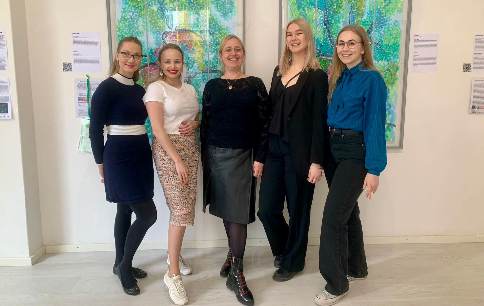
From left: Riina Harjumäki, Elle Koivunotko, Marjo Yliperttula, Noora Jansson, and Julia Monola from the Division of Pharmaceutical Biosciences, Drug Research Program, Faculty of Pharmacy, Helsinki University.
Professor Yliperttula and her research group focus on the creation of innovative biomaterials for cell culture, drug delivery, and tissue repair. These biomaterials are derived from nano-fibrillated cellulose, nanoparticles, and extracellular vesicles.
The research group has conducted successful pioneering research using Timegated® Raman spectroscopy including:
- Fluorescence-suppressed time-resolved Raman spectroscopy of pharmaceuticals using complementary metal-oxide-semiconductor (CMOS) single-photon avalanche diode (SPAD) detector
- Label-free characterization and real-time monitoring of cell uptake of extracellular vesicles
- Drug diffusivities in nanofibrillar cellulose hydrogel by combined time-resolved Raman and fluorescence spectroscopy
- Cellulase-assisted platelet-rich plasma release from nanofibrillated cellulose hydrogel enhances wound healing
Additionally, Marjo contributed/mentored Tatu Rojalin's and Jacopo Zini's the PhD thesis in which the new application of spectroscopy technique, in particular Timegated® (TG) Raman, in pharmaceutical and biopharmaceutical fields are investigated.
Table 1. The summary of publications related to time-gated Raman technology in the field of pharmaceuticals.
Fluorescence-suppressed Timegated® Raman spectroscopy for pharmaceuticals research
The first study evaluated the effectiveness of Timegated® Raman technology in suppressing fluorescence compared to traditional Raman spectroscopy. In the research, a short-wavelength, 532-nm picosecond pulsed laser coupled was utilized with a time-gated complementary metal-oxide-semiconductor (CMOS) single-photon avalanche diode (SPAD) detector to acquire Raman spectra of several drugs of interest. This was the first evidence of the penitential application of TG-Raman in the pharmaceutical field.
Raman spectroscopy is a highly advanced, sensitive, and user-friendly technology for biopharmaceutical measurements, Yliperttula describes.
Timegated® Raman spectroscopy enables fluorescence suppression and identification of the compounds
Compared to traditional Raman setups, the present Timegated® Raman technology has two major improvements. First, it is possible to overcome the strong fluorescence background that usually interferes with the much weaker Raman spectra. Second, using the high photon energy excitation light source, it is possible to generate a stronger Raman signal compared to traditional instruments.
Using Timegated® Raman, it was demonstrated for the first time the possibility of recording fluorescence-suppressed Raman spectra of solids, amorphous, crystalline, and non-photoluminescent and photoluminescent drugs such as caffeine, ranitidine hydrochloride, and amorphous and crystalline forms of indomethacin.
The data acquired by utilizing only the picosecond pulsed laser and a CMOS SPAD detector could be used for identifying the compounds directly without any data processing.
The value of Timegated® Raman for Pharmaceuticals, Process Analytical Technology (PAT) and Life Sciences
The obtained time-resolved Raman peaks were identified based on the calculations and existing literature. Raman spectra using non-time-resolved setups with continuous-wave excitation lasers were used as reference data. Overall, this demonstration of time-resolved Raman and fluorescence measurements with a CMOS SPAD detector shows promise in diverse areas, including fundamental chemical research, the pharmaceutical setting, process analytical technology (PAT), and the life sciences.

Studying extracellular vesicles with Timegated® Raman
Extracellular vesicles (EVs) are nanoparticles released by virtually all cell types in their environment. EVs are involved in cell-to-cell communications by transporting DNA, RNA, proteins, and lipids. They can also serve as molecular carriers and have potential applications in delivering drugs to target cells in various diseases. EVs, composed of particles in cellular membranes, are released into body fluids such as plasma, cerebrospinal fluid, semen, milk, and urine. The specific composition of EVs influences their function and interactions with cells, which in turn impacts the efficiency of their uptake by target cells.
Timegated® Raman technology contributes to the EVs research
In collaboration with the university, there were two studies conducted with the help of Timegated® Raman technology to show that Raman technology approaches contribute to a more comprehensive understanding of EVs and their applications in medical research and therapeutics.
Together, these studies underscore the versatility of EVs in medical research, emphasizing their potential to advance personalized medicine and targeted therapies.
EVs were studied from two different perspectives. The interest of the first study was to study label-free characterization and real-time monitoring of cell uptake of extracellular vesicles. The second study focused on the purity assessment of extracellular vesicles.
Time-resolved Raman enabled EV characterization and determination of the biochemical composition of the investigated EVs
The label-free approach for studying EVs was the characterization of EV composition by time-gated surface-enhanced Raman spectroscopy (TG-SERS). Time-gated SERS measurements were carried out using a PicoRaman spectrometer.
Previously the dynamics of EV uptake have been poorly characterized, mainly due to the scarcity of sensitive and dependable methods to monitor cellular uptake in real-time. With the help of time-gated Raman spectroscopy, it was possible to characterize the most abundant EVs of human blood, red blood cells (RBC)- and platelet (PLT)-derived EVs and study their interactions with prostate cancer cells. The TG-SERS spectra measured for RBC and PLT EVs in this study can be used for recognizing the most important molecular vibrations that differentiate these two EV types from each other.
The time-gated Raman instrument enabled suppression of the photo-degradation and autofluorescence typically present in biological samples and the time-gated Raman measurements revealed clear distinct features in the biochemical compositions of RBC and PLT EVs. Results revealed distinct biochemical features between the studied EVs and demonstrated that PLT-derived EVs were more efficiently internalized by PC-3 cells than RBC-derived EVs. Raman technology was found to efficiently complement conventional techniques and pave the way for further use of TG-SERS in EV studies.
Overall, time-gating enabled efficient suppression of photoluminescent background emission, typically prevalent in biological samples, such as EVs. TG-Raman can be efficiently used to reveal variabilities in the total protein, lipid, and cytosolic content of EVs of different origins. Furthermore, Raman spectroscopy shows broad potential for diagnostic purposes, for example, to investigate urinary EVs as biomarkers in diabetes.
In the pharmaceutical product manufacturing process, it is crucial to utilize label-free systems for accurate detection, and in this sense, Time-gated Raman is a phenomenal tool, Yliperttula praises.

Drug diffusivities and platelet-rich plasma released in nanofibrillar cellulose hydrogel
.jpg?width=1920&height=1080&name=hydrogel-illustration(1).jpg)
Hydrogels, of natural and synthetic origin, are actively studied for their use as implants and payload carriers. These biomaterials for delivery systems have enormous potential in basic biomedical research, drug development, and long-term delivery of biologics. To study the dynamics within hydrogel and its interaction with its payload is often challenging as sampling the hydrogel would alternate the condition of the complex hydrogel payload, which might affect the results of the measurements.
Spectroscopic methods could be ideal in this case, however, due to water-strong interference, IR is often ruled out, while UV spectroscopy often lacks resolution. The application of traditional Raman spectroscopy is often challenging due to fluorescence interference and low concentration of analytes. Here, using a long wavelength laser to overcome fluorescence interference might hamper the limit of detection and quantification of the technique. On the other hand, UV lasers might be too energetic and induce photodegradation or alteration of the sample.
Here, TG Raman, with its 532 laser and fluorescence suppression reveals an optimal choice. Additionally, TG Raman enables the simultaneous measurement of the Raman spectra, which contains information about the chemical nature of the samples, and the fluorescence lifetime (FLT). FLT is more sensitive than Raman to changes in the environment i.e. changes in viscosity and pH, this is particularly useful when we want to study the interaction between hydrogen and payload.
Particularly, we used TG Raman to study the diffusion of small drugs in NFC and the release rate of PRP – a source of growth factors, which are implicated in active tissue regeneration- from NFC in different conditions.
Apart from the technology, working with Timegate has also been extremely easy. Whenever we have had a scientific problem, there has always been a high-quality expert who has understood us and helped us move forward, Yliperttula notes appreciatively.


Sources used in this article:
Interviews with Marjo Yliperttula
The University of Helsinki, Research Units, Biopharmaceutics Group (https://researchportal.helsinki.fi/en/organisations/biopharmaceutics-group)
National Library of Medicine, Fluorescence-suppressed time-resolved Raman spectroscopy of pharmaceuticals using complementary metal-oxide semiconductor (CMOS) single-photon avalanche diode (SPAD) detector (https://pubmed.ncbi.nlm.nih.gov/26549117/)
ScienceDirect, Biosensors and Bioelectronics, Label-free characterization and real-time monitoring of cell uptake of extracellular vesicles (https://www.sciencedirect.com/science/article/abs/pii/S0956566320305029)
Journal of Controlled Release, Drug diffusivities in nanofibrillar cellulose hydrogel by combined time-resolved Raman and fluorescence spectroscopy (https://doi.org/10.1016/j.jconrel.2021.04.032)
Journal of Controlled Release, Cellulase-assisted platelet-rich plasma release from nanofibrillated cellulose hydrogel enhances wound healing (https://doi.org/10.1016/j.jconrel.2024.02.041)
University of Helsinki
The University of Helsinki is located in Finland. It is the oldest and largest institution of academic education in the country. The university is an international scientific community of 40,000 students and researchers. In international university rankings, the University of Helsinki typically ranks in the top 1 %. Through the power of knowledge, the University has contributed to society, education and welfare since 1640.