Timegated® Raman spectroscopy and proteomics analyses of hypoxic and normoxic renal carcinoma extracellular vesicles
An article Timegated® Raman spectroscopy and proteomics analyses of hypoxic and normoxic renal carcinoma extracellular vesicles is published in Nature - Scientific Reports.
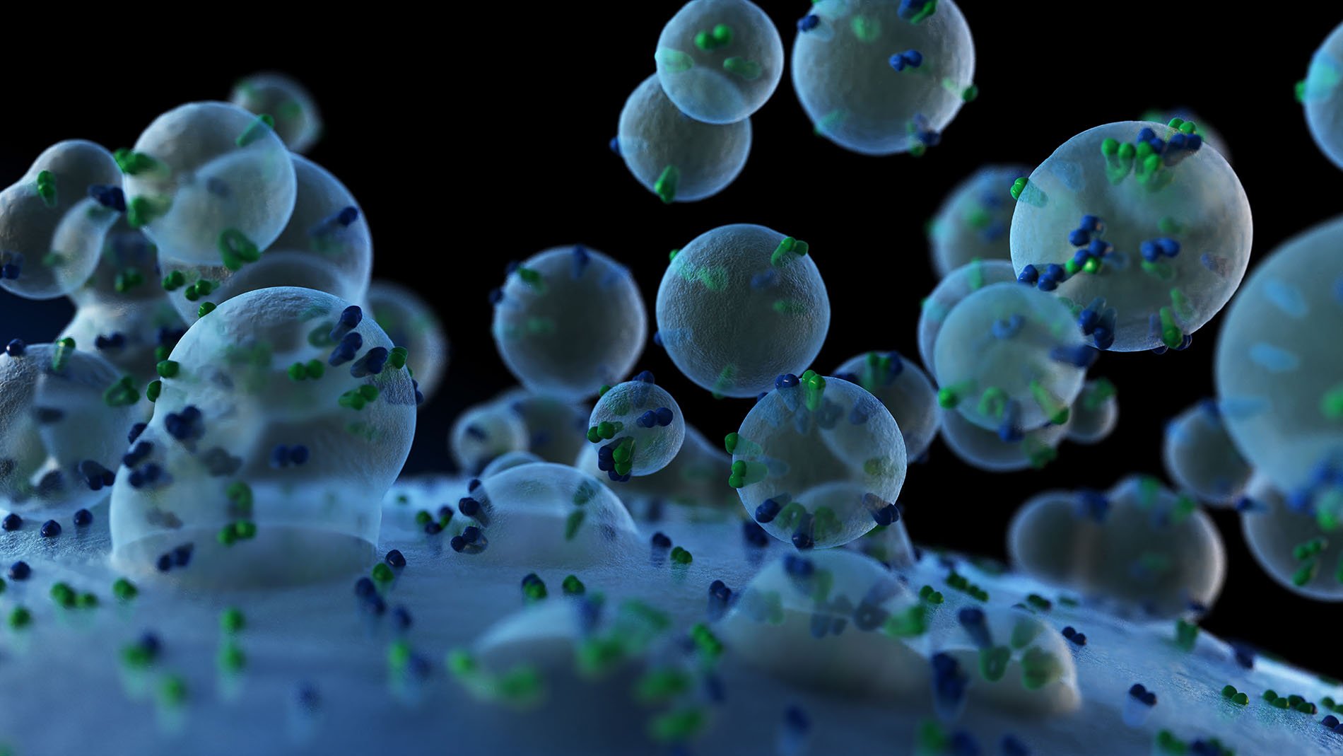
This publication highlights the important research topic of extracellular vesicles (EVs) which were studied using Timegated® Raman (TG-Raman) and surface-enhanced Timegated® Raman spectroscopy (TG-SERS). EVs are known to carry information from one cell to another and therefore it is currently a “hot topic” in cancer and biomedical research. Furthermore, EVs gained exponential interest because of their great potential to overcome many of the current obstacles that pharmaceutical industry is facing when searching for better drug delivery, therapeutic solutions, and novel biomarkers. Currently, EVs are an emerging business for drug delivery, diagnostic and analysis equipment. Unfortunately, novel characterization methods to develop these EVs, e.g. when purifying these vesicles, are still missing. Raman with SERS enhancement and without disturbing fluorescence is highly interesting since this serves as a potential technique to screen EVs even on the level of single EVs.
For the first time, with this paper the writers Anatoliy Samoylenko, Martin Kögler, Artem Zhyvolozhnyi, Olha Makieieva, Geneviève Bart, Sampson S. Andoh, Matthieu Roussey, Seppo J. Vainio & Jussi Hiltunen show that Timegated® Raman can distinguish between the low level of oxygen deficiency (hypoxia) and normoxia of EVs and that it is a fast and far less expensive method than mass-spectroscopy, showing comparable results. With TG-Raman, this article discloses even differences of EVs between mouse and human origin under the state of hypoxia and normoxia. Further the level of purity of EVs can be evaluated from EVs obtained from different protocols.
Read the open-access article.
Abstract
Extracellular vesicles (EVs) represent a diverse group of small membrane-encapsulated particles involved in cell–cell communication, but the technologies to characterize EVs are still limited. Hypoxia is a typical condition in solid tumors, and cancer-derived EVs support tumor growth and invasion of tissues by tumor cells. We found that exposure of renal adenocarcinoma cells to hypoxia induced EV secretion and led to notable changes in the EV protein cargo in comparison to normoxia. Proteomics analysis showed overrepresentation of proteins involved in adhesion, such as integrins, in hypoxic EV samples. We further assessed the efficacy of time-gated Raman spectroscopy (TG-RS) and surface-enhanced time-gated Raman spectroscopy (TG-SERS) to characterize EVs. While the conventional continuous wave excitation Raman spectroscopy did not provide a notable signal, prominent signals were obtained with the TG-RS that were further enhanced in the TG-SERS. The Raman signal showed characteristic changes in the amide regions due to alteration in the chemical bonds of the EV proteins. The results illustrate that the TG-RS and the TG-SERS are promising label free technologies to study cellular impact of external stimuli, such as oxygen deficiency, on EV production, as well as differences arising from distinct EV purification protocols.
If you would like to learn more about Timegted® technology, download Picoraman brochure from the link below:

