Biophotonics in biopharmaceutical applications with time-resolved Raman spectroscopy
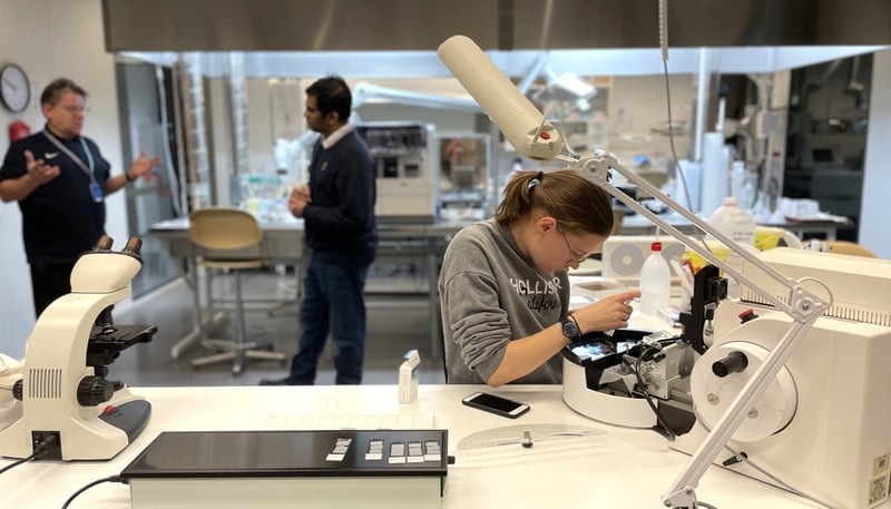
Timegate's new employee Dr. Jacopo Zini did his doctoral dissertation on biophotonics in biopharmaceutical applications. In his studies, he applied various photonics techniques in the characterization of complex biological samples. Among other techniques, he applied time-resolved Raman spectroscopy to study the interactions between drugs and biological material.
Read the doctoral thesis: Biophotonics in Biopharmaceutical Applications.
Abstract
Biopharmaceutical products are composed of complex or ordinate combinations of proteins, lipids, sugars, and nucleic acids or living cells or tissues. Due to the intrinsic variability of biological systems and the complexity of the bioprocesses involved in the production of these products, new technologies are required to monitor and characterize, the production processes and the final products. Biophotonic techniques, particularly Raman and Infrared (IR) spectroscopy are rapid, robust, operator independent, non-destructive and label free, thus particularly suitable for these purposes. This dissertation first investigates the use of biophotonic techniques in research of Extracellular Vesicles (EVs). EVs act as intercellular messengers and therefore have considerable potential in drug delivery system, diagnostic biomarker, or therapeutic agent. Subsequently highlighting the need and potential of a new kind of time gated Raman spectrometer to be created.
Raman and IR spectroscopy are methods able to characterize and assess the quality of EV suspensions with different degree of purity. These vibrational spectroscopic techniques show different intrinsic advantages, as they are label free and operator independent methods. Particularly, Raman might be the most suitable technology since it is less sensitive to water compared to IR. Raman spectroscopy reveals the information on the chemical composition and physical status of the analyte. However, it does not provide information of the analyte environment which the spectrofluorometer can gather. EVs studied by spectrofluorometer are required to be labelled with a fluorescent dye. Results obtained by fluorescence lifetime imaging spectroscopy underline that the cell uptake of the fluorescently labelled EVs is feasible. Attention must be paid to the efficacy of the labelling and further to the elimination of the unbound dye since the labelling may severely compromise the results or lead to wrong conclusions on EV functionality.
The combined advantage of Raman spectroscopy and fluorescence decay are obtained by a previously in-house developed time resolved Raman spectrometer. Thanks to the peculiar sensor of the spectrometer, the width of the time gate can be modified which is used to separate the Raman signal from the fluorescence tail. The modifications can be done even in the data post-processing phase, to obtain the best possible Raman signal-to-noise ratios. The simultaneously detected Raman spectra and time-resolved fluorescence decay curves are used to study the diffusion of small molecular drugs in a hydrogel. The data reveal the chemical composition, physical status, and the interaction with the environment of the samples. Taken together the obtained results suggest that the quality of EV suspensions can be assessed by Raman spectroscopy and their cell uptake detected by fluorescence lifetime spectroscopy. Both Raman spectra and fluorescence decay can be measured simultaneously by a second generation of time-resolved Raman spectrometer.
If you would like to learn more about Timegated® technology, download PicoRaman M3 brochure here.