Studies of Timegated® Raman in Life Sciences
Have you explored any scientific publications for applying time-gated Raman in life sciences? Now you have the chance, as we are introducing a review article about the principle of the technology and three fascinating peer-reviewed articles demonstrating time-gated Raman technology as a valuable tool in life science applications such as escherichia coli cultivation and extracellular vesicle discrimination.
If you have not seen our last blog, you can find it here: Top 8 benefits of Timegated® Raman in process monitoring.
Time-gated Raman spectroscopy principle, history, and future developments
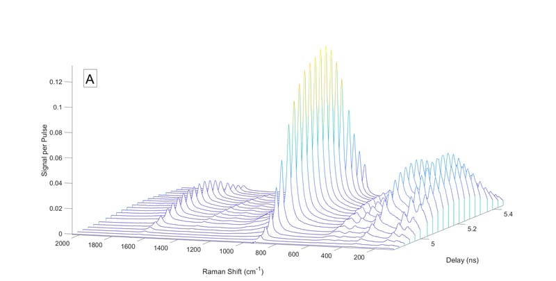
Martin Kögler’s and Bryan Heilala’s article ”Time-gated Raman spectroscopy – a review” in Measurement Science and Technology (IOPscience) introduces the working principle, history, and future prospects of time-gated Raman spectroscopy development and applications.
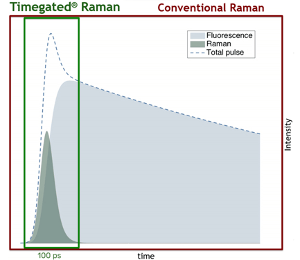
Graph demonstrating the Timegated® Raman breakthrough innovation. Pulsed picosecond range lasers make it possible to acquire Raman spectra with real fluorescence suppression.
Abstract
Time-gated (TG) Raman spectroscopy has been shown to be an effective technical solution for the major problem of sample-induced fluorescence, masking Raman signal during spectral detection. Technical methods of fluorescence rejection have come a long way from the early implementations of large and expensive laboratory equipment, such as the optical Kerr-gate. Today there are some more affordable small sized options available. These improvements are largely due to advances in the production of spectroscopic and electronic components that further led to the reduction of device complexity and costs. An integral part of TG Raman is the temporally precise synchronization (picosecond range) between the pulsed laser excitation source and the sensitive and fast detector. The detector is able to collect the Raman signal during the short laser pulses while fluorescence emission, which has a longer delay, is rejected during the detector dead-time. TG Raman is also resistant against ambient light as well as thermal missions due to the short measurement duty cycle. In recent years, the focus of ultra-sensitive and fast detectors has been on gated and intensified charge coupled devices (ICCDs) or on CMOS single-photon avalanche diode (SPAD) arrays, which are also suitable for performing TG Raman spectroscopy. SPAD arrays have the advantage of being even more sensitive with better temporal resolution compared to gated CCDs without the necessity of excessive detector cooling. This review aims to provide an overview of TG Raman from early to recent developments, applications and extensions.
Timegated® Raman assessing recombinant protein production and monitoring escherichia coli cultivation
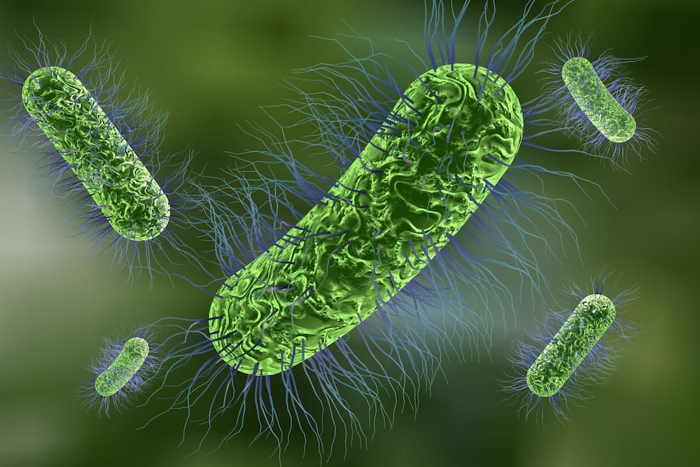 Escherichia coli bacteria.
There is a broad range of applications for recombinant proteins ranging from research to pharmaceutical development. Bacteria have been extensively used to produce recombinant proteins. Due to the inherent challenges of the process, monitoring is vital. In this regard, time-gated surface-enhanced Raman spectroscopy (TG-SERS) was utilized to assess recombinant protein production in Escherichia coli. Kögler et al. (2018) monitored metabolite concentrations of Escherichia Coli bioreactor cultivations in cell-free supernatant samples. They investigated the concentrations of glucose, acetate, AMP, and cAMP. Further, the same group employed time-gated Raman spectroscopy and showed that this technology is a powerful tool to evaluate protein production and correct folding within living E. coli cells during cultivation. To further learn about how Timegated® Raman spectroscopy was used for this application, you can explore the following articles:
Kögler M, Itkonen J, Viitala T, Casteleijn M G: Assessment of recombinant protein production in E. coli with Time-Gated Surface Enhanced Raman Spectroscopy (TG-SERS) in Scientific Reports.
Abstract
Time-Gated Surface-Enhanced Raman spectroscopy (TG-SERS) was utilized to assess recombinant protein production in Escherichia coli. TG-SERS suppressed the fluorescence signal from the biomolecules in the bacteria and the culture media. Characteristic protein signatures at different time points of the cell cultivation were observed and compared to conventional continuous wave (CW)-Raman with SERS. TG-SERS can distinguish discrete features of proteins such as the secondary structures and is therefore indicative of folding or unfolding of the protein. A novel method utilizing nanofibrillar cellulose as a stabilizing agent for nanoparticles and bacterial cells was used for the first time in order to boost the Raman signal, while simultaneously suppressing background signals. We evaluated the expression of hCNTF, hHspA1, and hHsp27 in complex media using the batch fermentation mode. HCNTF was also cultivated using EnBase in a fed-batch like mode. HspA1 expressed poorly due to aggregation problems within the cell, while hCNTF expressed in batch mode was correctly folded and protein instabilities were identified in the EnBase cultivation. Time-gated Raman spectroscopy showed to be a powerful tool to evaluate protein production and correct folding within living E. coli cells during the cultivation.
Kögler M, Paul A, Anane E, Birkholz M, Bunker A, Viitala T, Maiwald M, Junne S & Neubauer P: Comparison of time-gated surface-enhanced raman spectroscopy (TG-SERS) and classical SERS based monitoring of Escherichia coli cultivation samples in Biotechnology Progress (AIChE).
Abstract
The application of Raman spectroscopy as a monitoring technique for bioprocesses is severely limited by a large background signal originating from fluorescing compounds in the culture media. Here, we compare time-gated Raman (TG-Raman)-, continuous wave NIR-process Raman (NIR-Raman), and continuous wave micro-Raman (micro-Raman) approaches in combination with surface enhanced Raman spectroscopy (SERS) for their potential to overcome this limit. For that purpose, we monitored metabolite concentrations of Escherichia coli bioreactor cultivations in cell-free supernatant samples. We investigated concentration transients of glucose, acetate, AMP, and cAMP at alternating substrate availability, from deficiency to excess. Raman and SERS signals were compared to off-line metabolite analysis of carbohydrates, carboxylic acids, and nucleotides. Results demonstrate that SERS, in almost all cases, led to a higher number of identifiable signals and better resolved spectra. Spectra derived from the TG-Raman were comparable to those of micro-Raman resulting in well-discernable Raman peaks, which allowed for the identification of a higher number of compounds. In contrast, NIR-Raman provided a superior performance for the quantitative evaluation of analytes, both with and without SERS nanoparticles when using multivariate data analysis.

Timegated® Raman monitoring the discrimination of extracellular vesicles
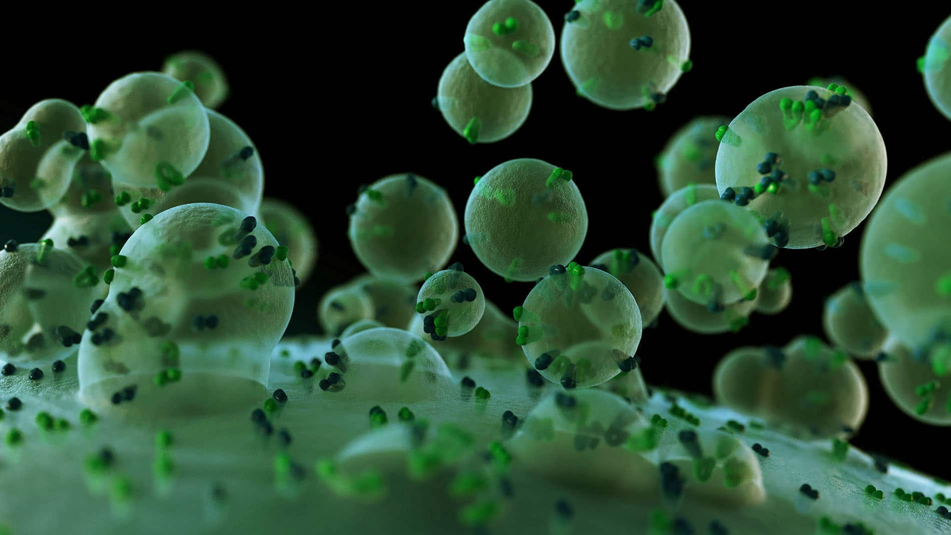 Extracellular vesicles.
Owing to the versatility of the Time-gated Raman technology, it can be employed to diagnose diseases and test therapeutic prognosis. Samoylenko et al. have studied both the hypoxic and normoxic extracellular vesicles (EVs) from renal carcinoma cell lines. The last decade has attracted a lot of interest in research concerning EVs. These vesicles typically contain various proteins, RNA species, and metabolites. The composition of the EVs reflects the physiological status of a cell that produces, providing ground to develop novel diagnostic tools. In this study, the authors have endeavored to discriminate between the two groups of vesicles. Time-gated Raman has proved to be a useful, reliable label-free, and fast technique to this end. To learn more about this latest research please read:
Samoylenko A, Kögler M, Zhyvolozhnyi A, Makieieva O, Bart G, Andoh S S, Roussey M, Vainio S J, Hiltunen J: Time-gated Raman spectroscopy and proteomics analyses of hypoxic and normoxic renal carcinoma extracellular vesicles in Scientific Reports).
Abstract
Extracellular vesicles (EVs) represent a diverse group of small membrane-encapsulated particles involved in cell–cell communication, but the technologies to characterize EVs are still limited. Hypoxia is a typical condition in solid tumors, and cancer-derived EVs support tumor growth and invasion of tissues by tumor cells. We found that exposure of renal adenocarcinoma cells to hypoxia induced EV secretion and led to notable changes in the EV protein cargo in comparison to normoxia. Proteomics analysis showed overrepresentation of proteins involved in adhesion, such as integrins, in hypoxic EV samples. We further assessed the efficacy of time-gated Raman spectroscopy (TG-RS) and surface-enhanced time-gated Raman spectroscopy (TG-SERS) to characterize EVs. While the conventional continuous wave excitation Raman spectroscopy did not provide a notable signal, prominent signals were obtained with the TG-RS that were further enhanced in the TG-SERS. The Raman signal showed characteristic changes in the amide regions due to alteration in the chemical bonds of the EV proteins. The results illustrate that the TG-RS and the TG-SERS are promising label free technologies to study cellular impact of external stimuli, such as oxygen deficiency, on EV production, as well as differences arising from distinct EV purification protocols.
If you have any questions regarding Timegated® Raman spectroscopy, applications, or our instrument, please contact us.
Stay tuned for more about Timegated® Raman and EVs!